Retinal Vein Occlusion
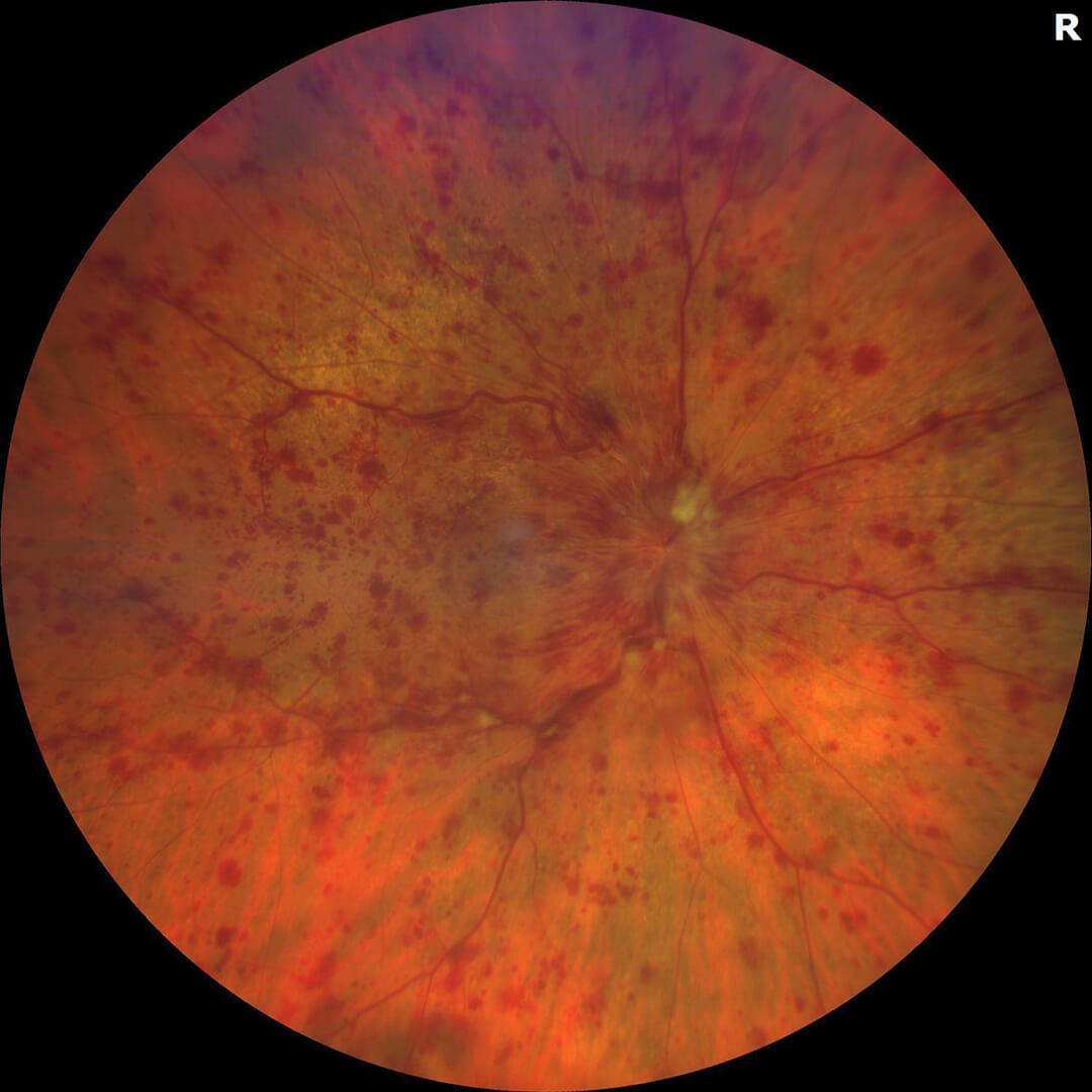
Diffuse retinal hemorrhage, or bleeding, from a retinal vein occlusion.
The function of blood cells is to deliver oxygen to our tissues such as muscle, skin, brain and eyes in order to function properly. When blood flow is blocked the tissue in question begins to malfunction. In the case of a retinal vein occlusion, blockage of a retinal vein causes poor blood flow resulting in damage to the retina and poor vision. When the main retinal vein is blocked it is called a central retinal vein occlusion (CRVO). If a smaller retinal vein is blocked it is called a branch retinal vein occlusion (BRVO). When either retinal vein becomes blocked, blood collects in the retinal tissue resulting in retinal hemorrhages, leakage and swelling of the retina called macular edema.
Risk factors that may increase your risk for a retinal vein occlusion include high blood pressure, diabetes, vascular disease, and glaucoma.
Retinal vein occlusion almost always occurs in one eye but when it is present in both eyes we worry about diseases that cause a general “thickening” of the blood. These conditions can lead to blood clots. In such cases we recommend more extensive blood testing, evaluation by your primary care physician and sometimes a referral to a hematologist.
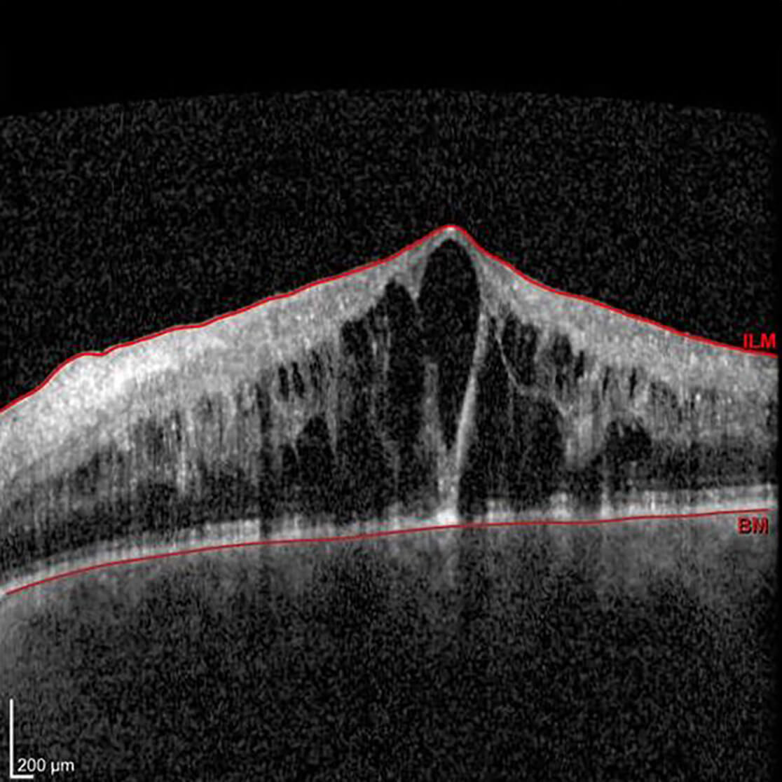
3D scans of the macula show accumulated fluid, or macular edema as cystic black spaces resulting in thickening of the tissue
There is no cure for retinal vein occlusions but there are treatments for some of the sight threatening complications of retinal vein occlusion. Most frequently retinal vein occlusions are treated with anti-VEGF medications (link to article). Other treatments include steroid injections and laser, although these are used less commonly.